Home

Egomanija Kreta Trebuh mcherry excitation laser confocal Mlečni izdelki Gnezdo Rama

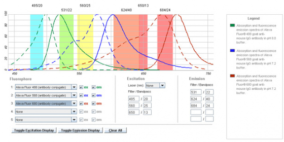
Fluorophore – The Live Cell Imaging facility

Confocal spectral microscopy, a non-destructive approach to follow  contamination and biofilm formation of mCherry Staphylococcus aureus on  solid surfaces | Scientific Reports
Confocal spectral microscopy, a non-destructive approach to follow contamination and biofilm formation of mCherry Staphylococcus aureus on solid surfaces | Scientific Reports
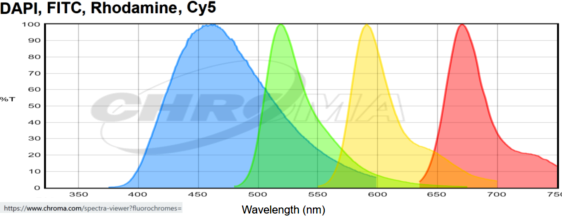
Spectra of common fluorophores | Light Microscopy Core Facility

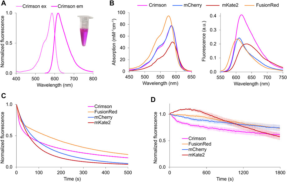
Frontiers | A Bright, Nontoxic, and Non-aggregating red Fluorescent Protein  for Long-Term Labeling of Fine Structures in Neurons
Frontiers | A Bright, Nontoxic, and Non-aggregating red Fluorescent Protein for Long-Term Labeling of Fine Structures in Neurons

FITC and mCherry form a suitable pair of fluorochromes for two-color... |  Download Scientific Diagram
FITC and mCherry form a suitable pair of fluorochromes for two-color... | Download Scientific Diagram

Confocal spectral microscopy, a non-destructive approach to follow  contamination and biofilm formation of mCherry Staphylococcus aureus on  solid surfaces | Scientific Reports
Confocal spectral microscopy, a non-destructive approach to follow contamination and biofilm formation of mCherry Staphylococcus aureus on solid surfaces | Scientific Reports
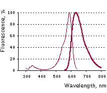
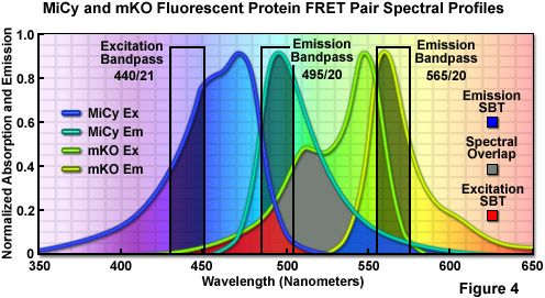
Evrogen mKate2: Detailed description

Choosing Your Fluorescent Proteins for Multi-Color Imaging

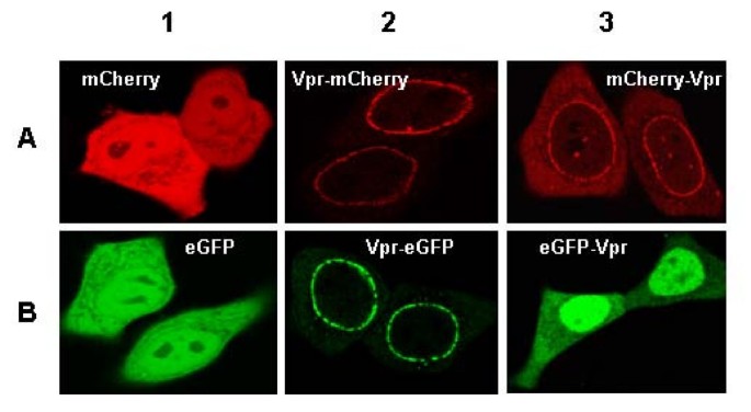
Direct Vpr-Vpr Interaction in Cells monitored by two Photon Fluorescence  Correlation Spectroscopy and Fluorescence Lifetime Imaging | Retrovirology  | Full Text
Direct Vpr-Vpr Interaction in Cells monitored by two Photon Fluorescence Correlation Spectroscopy and Fluorescence Lifetime Imaging | Retrovirology | Full Text

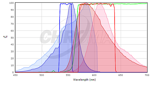
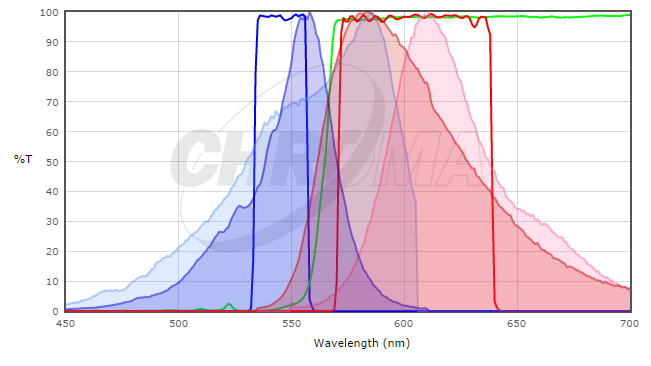
The filter settings for visualization of GFP and mCherry fluorescence... |  Download Scientific Diagram
The filter settings for visualization of GFP and mCherry fluorescence... | Download Scientific Diagram

Red fluorescent proteins for imaging Zymoseptoria tritici during invasion  of wheat - ScienceDirect
Red fluorescent proteins for imaging Zymoseptoria tritici during invasion of wheat - ScienceDirect

Visualization of MPC mCherry spheroids using confocal laser scanning... |  Download Scientific Diagram
Visualization of MPC mCherry spheroids using confocal laser scanning... | Download Scientific Diagram

Confocal laser scanning microscopy of cells co-expressing (A) EGFP-Bak... |  Download Scientific Diagram
Confocal laser scanning microscopy of cells co-expressing (A) EGFP-Bak... | Download Scientific Diagram

Confocal Microscopy - Fluorophores for Confocal Microscopy | Olympus LS

Choosing Your Fluorescent Proteins for Multi-Color Imaging

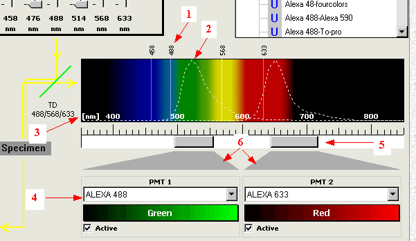
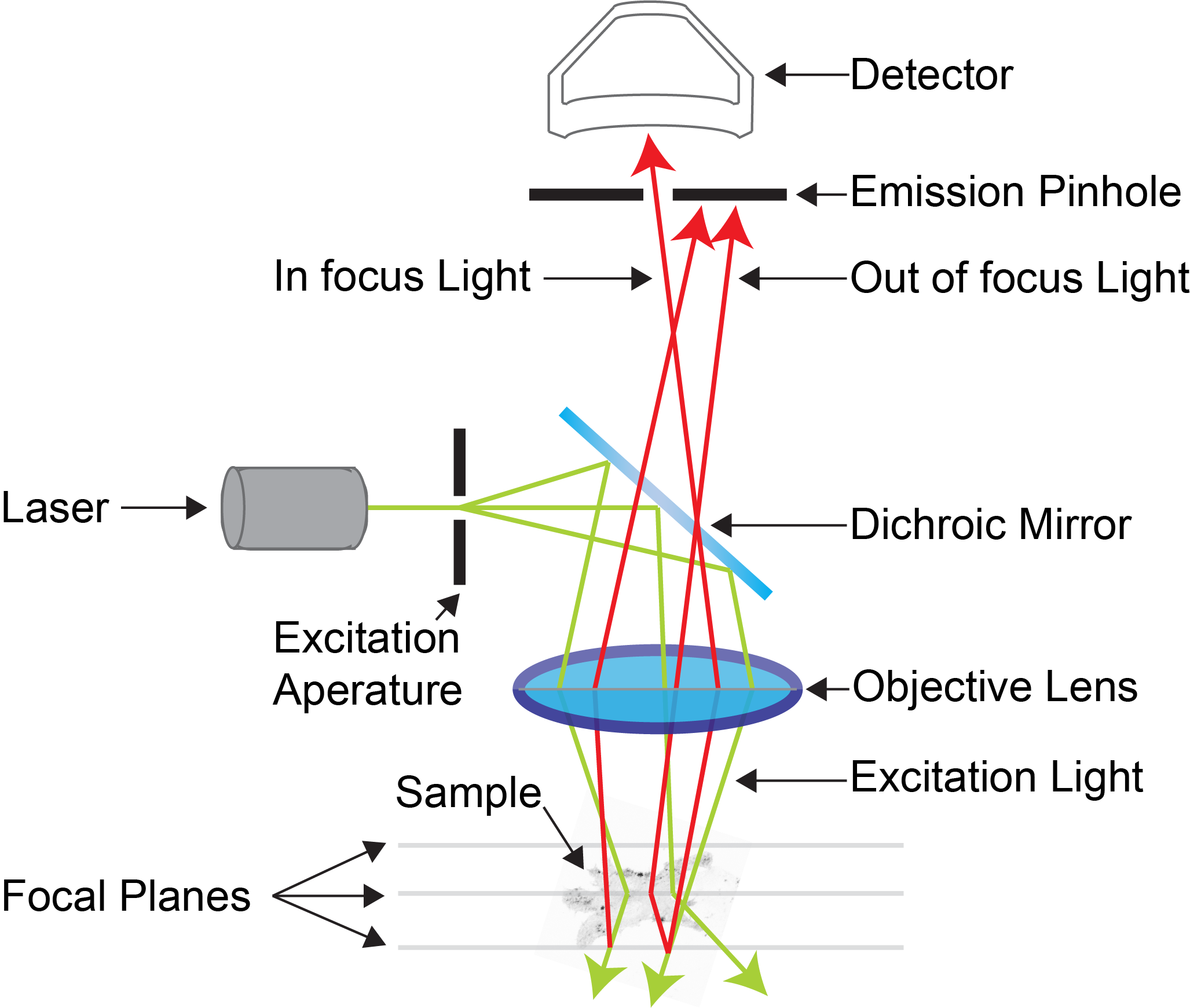
Confocal tutorial Optimization: emission collecting. AMU Dept Pathol Univ  Hel
Confocal tutorial Optimization: emission collecting. AMU Dept Pathol Univ Hel

Conformational Dynamics of mCherry Variants: A Link between Side-Chain  Motions and Fluorescence Brightness | The Journal of Physical Chemistry B
Conformational Dynamics of mCherry Variants: A Link between Side-Chain Motions and Fluorescence Brightness | The Journal of Physical Chemistry B

A guide to choosing fluorescent protein combinations for flow cytometric  analysis based on spectral overlap - Kleeman - 2018 - Cytometry Part A -  Wiley Online Library
A guide to choosing fluorescent protein combinations for flow cytometric analysis based on spectral overlap - Kleeman - 2018 - Cytometry Part A - Wiley Online Library

Detecting Protein Complexes in Living Cells from Laser Scanning Confocal  Image Sequences by the Cross Correlation Raster Image Spectroscopy Method:  Biophysical Journal
Detecting Protein Complexes in Living Cells from Laser Scanning Confocal Image Sequences by the Cross Correlation Raster Image Spectroscopy Method: Biophysical Journal

Chemosensors | Free Full-Text | Real-Time Fluorescence Imaging of  His-Tag-Driven Conjugation of mCherry Proteins to Silver Nanowires
Chemosensors | Free Full-Text | Real-Time Fluorescence Imaging of His-Tag-Driven Conjugation of mCherry Proteins to Silver Nanowires

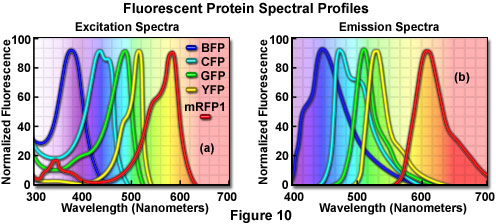
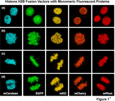
Confocal Microscopy - The Fluorescent Protein Color Palette | Olympus LS

fluorescent probes for microscopy

Confocal Microscopes - Institute for Molecular Bioscience - University of  Queensland
Confocal Microscopes - Institute for Molecular Bioscience - University of Queensland

Confocal Microscopy - The Fluorescent Protein Color Palette | Olympus LS

The filter settings for visualization of GFP and mCherry fluorescence... |  Download Scientific Diagram
The filter settings for visualization of GFP and mCherry fluorescence... | Download Scientific Diagram
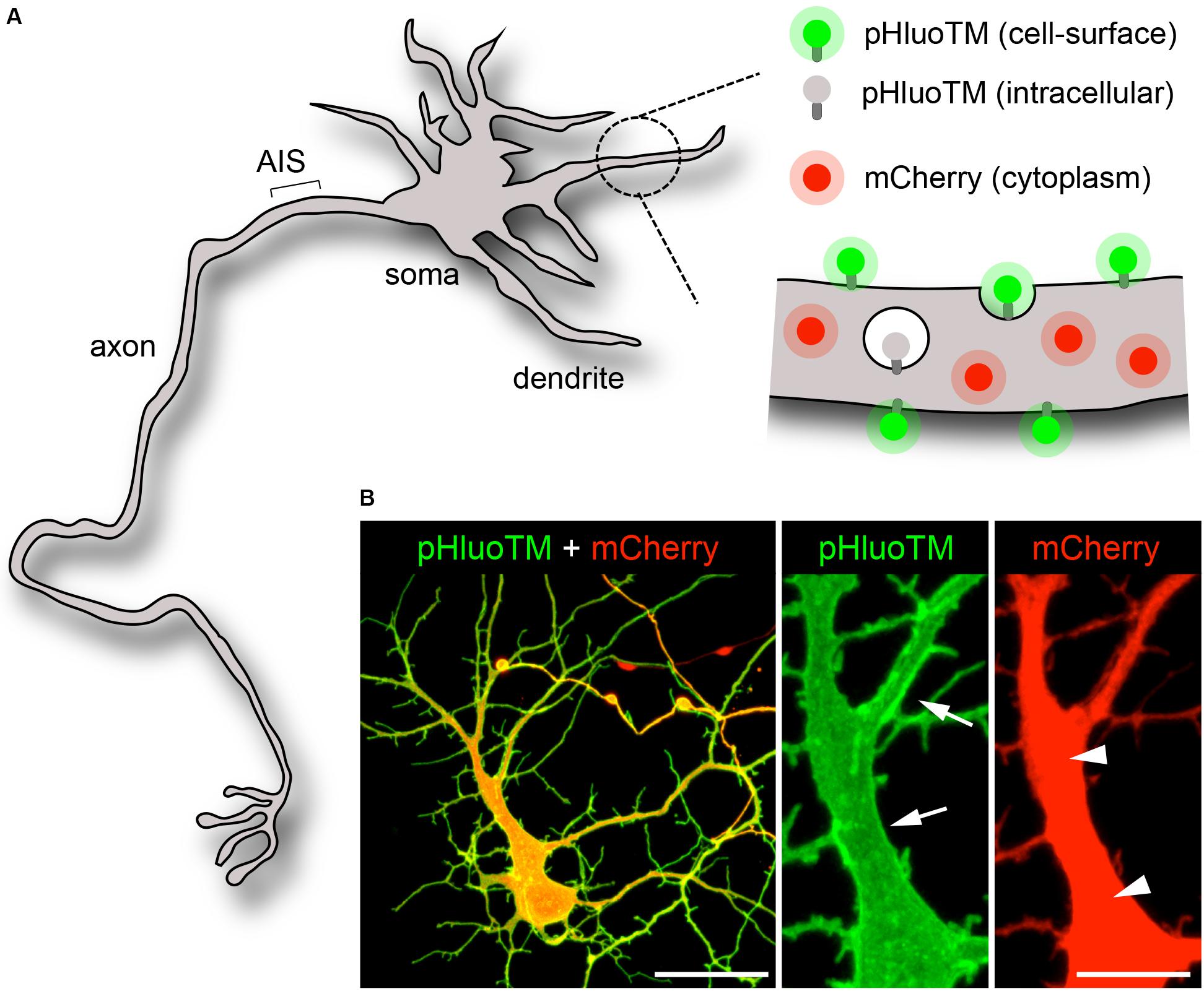
Frontiers | Whole-Cell Photobleaching Reveals Time-Dependent  Compartmentalization of Soluble Proteins by the Axon Initial Segment
Frontiers | Whole-Cell Photobleaching Reveals Time-Dependent Compartmentalization of Soluble Proteins by the Axon Initial Segment